
Anterior cervical fusion (ACF) with autologous bone has been reported over 50 years ago. The continuous development of materials with elastic modulus close to that of the cortical bone improves induction of osteogenesis and simplifies the technique of interbody fusion.
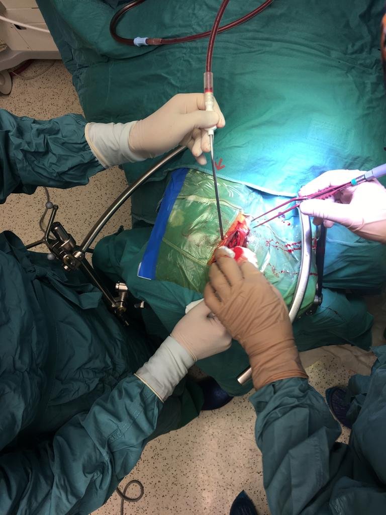
Anterior cervical microdiscectomy is usually performed for degenerative disk disease (DDD) that produces osteophytes and/or herniated disk provoking radiculopathy and/or myelopathy. Surgical intervention is indicated after failure of adequate conservative treatment. When spinal cord compression for cervical DDD at one or more disc levels causes myelopathy with or without brachialgia, anterior cervical microdiscectomy and osteophytectomy followed by ACF are almost worldwide accepted standard procedures, even if several Authors sustain that fusion procedure following anterior cervical discectomy is not necessary.
The goals of bone substitutes for ACF are: biomechanical support, restore foraminal height, shore up cervical lordosis, optimal osteointegration, reduce donor site complication, shorter hospital stay. During the last two decades stand alone cages of various materials, allowing immediate stability without plates and screws, have been developed.
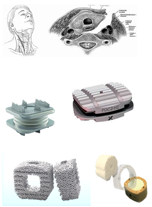
The ideal cage for ACF should ensure immediate structural support and subsequent adequate osteointegration and stability. A determining factor is the capacity of the material to be elastically deformed. This capacity is the elastic modulus (EM): higher EM requires higher forces for deforming temporarily the material. Among several metals, TM (porous tantalum) is an open-cell porous biomaterial with a structure similar to trabecular bone. The structure of porous tantalum affords a high volumetric porosity, a low EM, and relatively high frictional characteristics. Moreover, TM is inert and resistant to acid corrosion. All these characteristics make tantalum ideal for orthopedic implants and, therefore, an excellent biomaterial for ACF cages.
On the basis of these considerations, we investigated the bony incorporation of TM cages inserted in the interspace after anterior cervical spine microdiscectomy and osteophytectomy. Outcomes, fusion rates, and complications were assessed in 88 consecutive patients with a minimum follow-up of 12 months operated on for cervical DDD producing one- or two-level spondylotic myelopathy.